 |
 |
The ΔNp63 isoform, also known as p40, is the predominant isoform of p63 that is truncated, or lacking the N-terminal domain. p40 is a nuclear protein and a transcription factor. It is confined to basal cells
of squamous epithelia and urothelium, as well as basal cells/myoepithelial cells in breast, sweat gland, salivary gland, and prostate. Recent studies have shown that p40 is highly specific for squamous and basal cells and is superior to p63 for diagnosing lung squamous cell carcinoma.
Napsin A is a protease that is predominantly expressed in the lung and kidney. It is expressed in alveolar type II cells. Napsin A is detected in 60-90% of nonmucinous lung adenocarcinoma and less frequently in mucinous lung adenocarcinoma and large cell carcinoma (20-30%). In most studies, Napsin A is not detected, or only focally staining, in lung squamous cell carcinoma. Studies have shown that Napsin A has approximately the same sensitivity as TTF1, but specificity is higher
Intended use
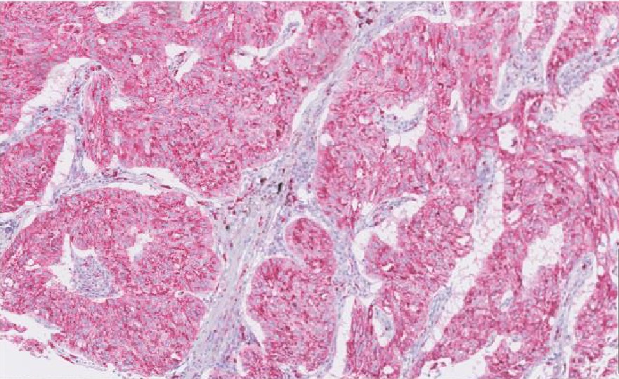
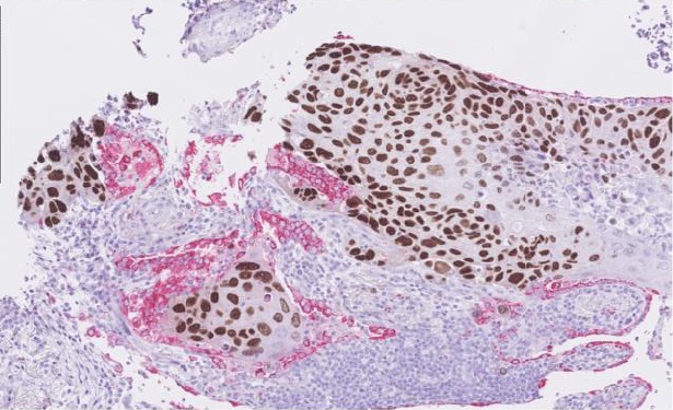
The DUO anti-p40 [BC28] / Napsin A [EP205] Antibody Cocktail utilizes the brown DAB chromogen for nuclear p40 and the AP Red chromogen for cytoplasmic Napsin A. The antibody cocktail is a useful aid in differentiating lung squamous cell carcinoma (most often p40 positive and Napsin A negative) from lung adenocarcinoma (most often p40 negative and Napsin A positive) when used with a panel of other antibodies
|
Specifications |
|
|
product code: 8487-C010 |
Clone: BC28 / EP205 |
|
Staining pattern: Nuclear staining (p40, DAB), cytoplasmic staining (Napsin A, RED) |
Control tissue: Tonsil, lung, placenta, kidney, appendix |
|
Associated panels: Lung panel |
|
|
Assessment criteria |
no scan available at this time |
|
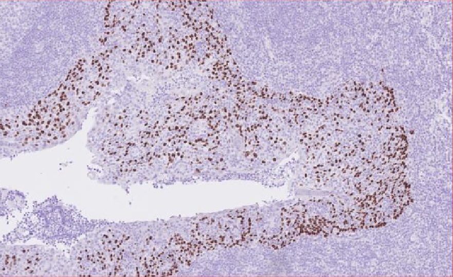
A moderate to strong, distinct nuclear staining reaction of virtually all squamous epithelial cells in the tonsil (p40) |
 |
|
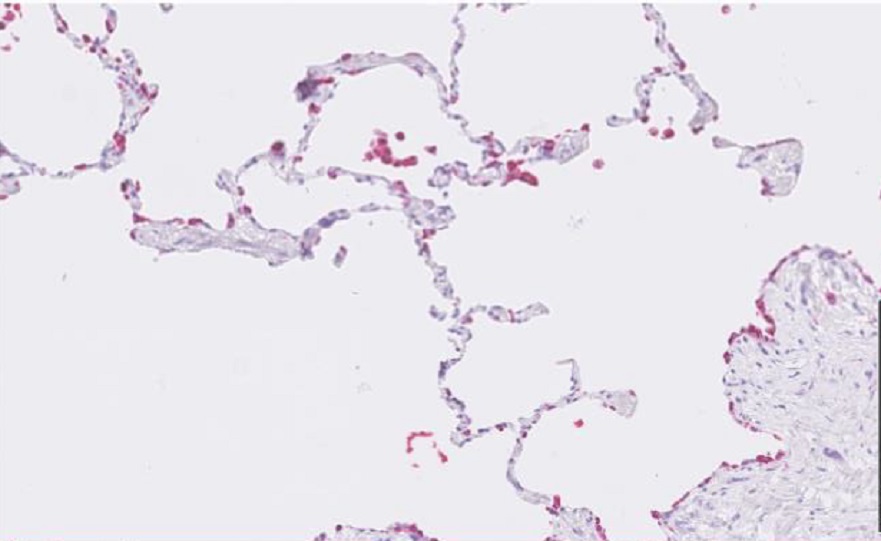
Virtually all type II pneumocytes and alveolar macrophages in lung must show a moderate to strong, granular cytoplasmic staining reaction (Napsin A) |
 |
|
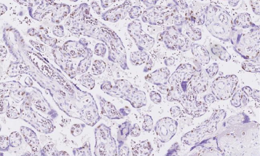
An at least weak to moderate, distinct nuclear staining reaction of cytotrophoblasts in placenta must be seen (p40) |
 |
| Virtually all epithelial cells of the proximal tubules in kidney must show an at least moderate, granular cytoplasmic staining reaction for Napsin A (no ideal tissue with low level expression being identified) | 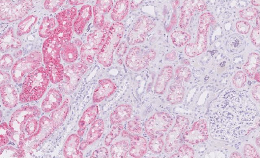 |
| No staining reaction should be seen including lymphocytes and epithelial cells in appendix (p40 and Napsin A) - (colon shown here..) | 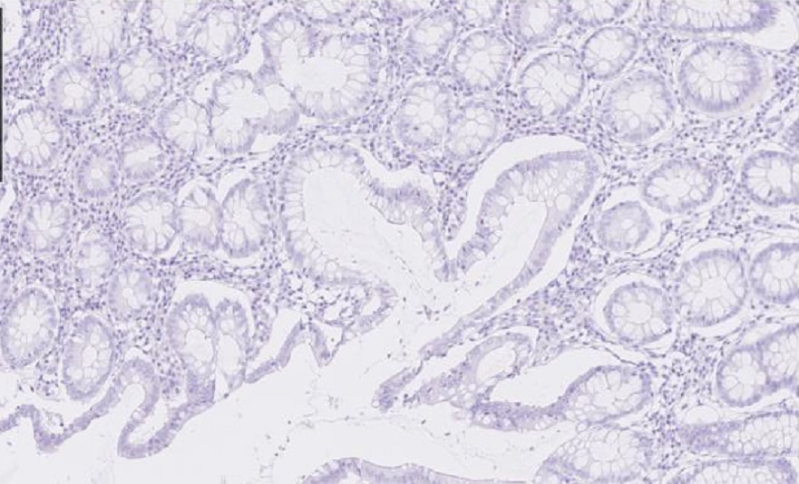 |
| example: lung adenoca. | example: lung squamous cell ca. |
 |
 |
| Related articles: | |
Best Practices Recommendations for Diagnostic Immunohistochemistry in Lung Cancer. J Thorac Oncol. 2019 Mar;14(3):377-407 |
|
| Use of dual-marker staining to differentiate between lung squamous cell carcinoma and adenocarcinoma. J Int Med Res. 2020 Apr;48(4) | |

Comments
0 comments
Please sign in to leave a comment.